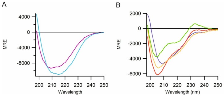
Figure 4.
Far-ultraviolet circular dichroism (CD) spectra for the purified proteins. (A) The spectra of mucin 2 (V36-G389; light blue) and alpha tectorin (P707-P981; purple). (B) Spectra of constructs of region 3 of perlecan; G503-Q874 (dark blue), S762-R1158 (red), R1158-T1672 (green) and G503-T1672 (orange). All constructs show spectra consistent with their expected natively folded state at near-physiological ionic strength.

