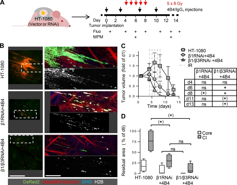
Figure 5.
Radiosensitization of HT-1080 tumors by β1/β3 integrin RNA interference combined with antibody-based β1 integrin targeting. (A) Protocol for administration of anti-β1 (4B4) or IgG1 combined with fractionated IR and sequential intravital imaging of the tumor response. Fluo, epifluorescence overview microscopy. MPM, subcellular-resolved multiphoton microscopy. (B) Topology and extent of the invasion zone in response to fractionated IR combined with single-integrin (β1) or dual β1/β3 integrin interference. Epifluorescence (left) and 3D reconstructed z-projections from regions marked by dashed boxes using multiphoton microscopy (right; day 13). White asterisks, apoptotic nuclei. Scale bars, 1 mm (left); 250 µm (right). (C) Time-dependent tumor volume. Data show the means ± SD from three to four independent tumors, with P values for comparing irradiated integrin-targeted tumors to irradiated control tumors. (*), P = 0.006; *, P = 0.004. Statistics, Mann–Whitney U test (Bonferroni-corrected threshold: P = 0.005). (D) Regression of tumor core and collective invasion (CI) zone after IR with or without integrin mono- or dual interference. Data show median residual areas, 25th/75th percentiles (box), and 5th/95th percentiles (whiskers) of day 13 normalized to day 6. Per condition, four tumors were analyzed. (*), P = 0.03; ns, not significant. Statistics, Mann–Whitney U test (Bonferroni-corrected threshold: P = 0.0125).

