Figure S1.
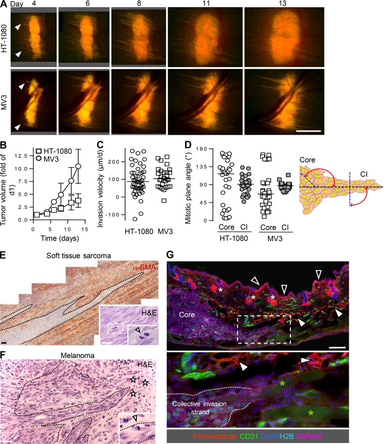
Kinetics and organization of collective invasion in mouse dermis and comparison to human samples. (A and B) Time-dependent whole-tumor morphology (A) and tumor volume (B) of HT-1080 and MV3 xenografts in the skin window. Data represent the means and SD from five (HT-1080) and three (MV3) tumors. Arrowheads, onset of collective invasion. Scale bar, 1 mm. (C) Median velocity of collective invasion into the dermis. Data represent the distance migrated per day from day 4 to 6 of 48 (HT-1080) and 28 (MV3) individual collective strands from three (HT-1080) and two (MV3) tumors. Negative values originate from occasional rearward orientation or retraction of strand tips. (D) Orientation of mitotic planes in the core or collective invasion (CI) strands relative to the direction of migration. A median angle of ∼90° reflects mitotic planes aligned perpendicular to the invasion direction. Per region and tumor type, ∼30 mitotic planes were analyzed from one tumor. (E) Collective invasion pattern in human adult primary soft tissue sarcoma in subdiaphragmal location. Multicellular strands bordered by reactive α-smooth muscle actin (SMA)–positive stromal cells. Inset, mitotic planes (arrowhead) orientated perpendicularly to the invasion direction. Dashed lines, border between invasion zone and stroma. Scale bars, 100 µm. (F) Collective invasion pattern in human primary melanoma lesion during vertical growth phase with deep dermal invasion. Inset, mitotic plane (arrowhead) perpendicular to invasion direction. Dashed lines, border stroma to invasion zone. Asterisks, individual tumor cells. Scale bar, 100 µm. (G) Absence of hypoxic areas in HT-1080 xenograft after 7 d of growth in the skin window. Hypoxic regions detected by pimonidazole staining (red) include the upper epidermis (black arrowheads), sebaceous glands (white asterisks), and dermal fat tissue (white arrowheads), but not the tumor core. Green asterisks, autofluorescent myofibers. Scale bar, 1 mm.

