Figure S1.
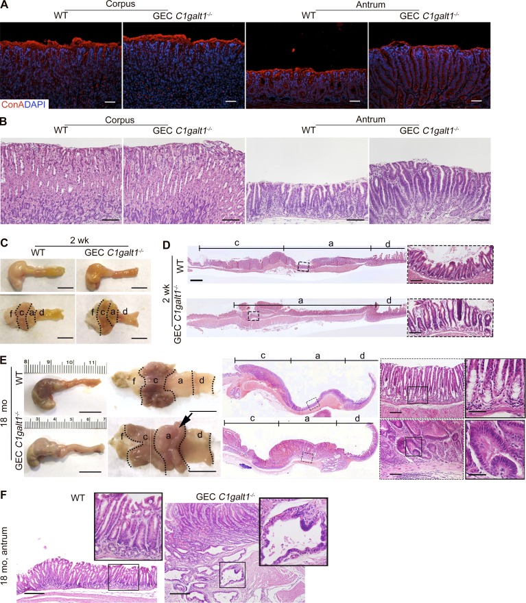
GEC C1galt1−/− mice develop spontaneous gastritis at an early age and gastric cancer at age 18 mo. (A) Representative images of IF-stained WT and GEC C1galt1−/− stomach sections. (B) H&E staining of corpus and antrum. (C) Representative gross morphology of murine stomach (n = 4–5 mice/group). Scale bars, 1 cm. a, antrum; c, corpus; d, duodenum; f, forestomach. (D) H&E staining of stomach sections. Inset: Magnified portion of boxed region in left image. Scale bars, 200 µm (tiling images); 50 µm (inset). (E) Representative gross morphology of murine stomach (n = 4–5 mice/group). Arrow shows a large antral tumor. Scale bars, 1 cm (gross morphology); 200 µm (tiling images); 50 µm (left inset); 25 µm (right inset). (F) H&E staining of antrum. Inset: Magnified image of boxed region. Scale bars, 100 µm.

