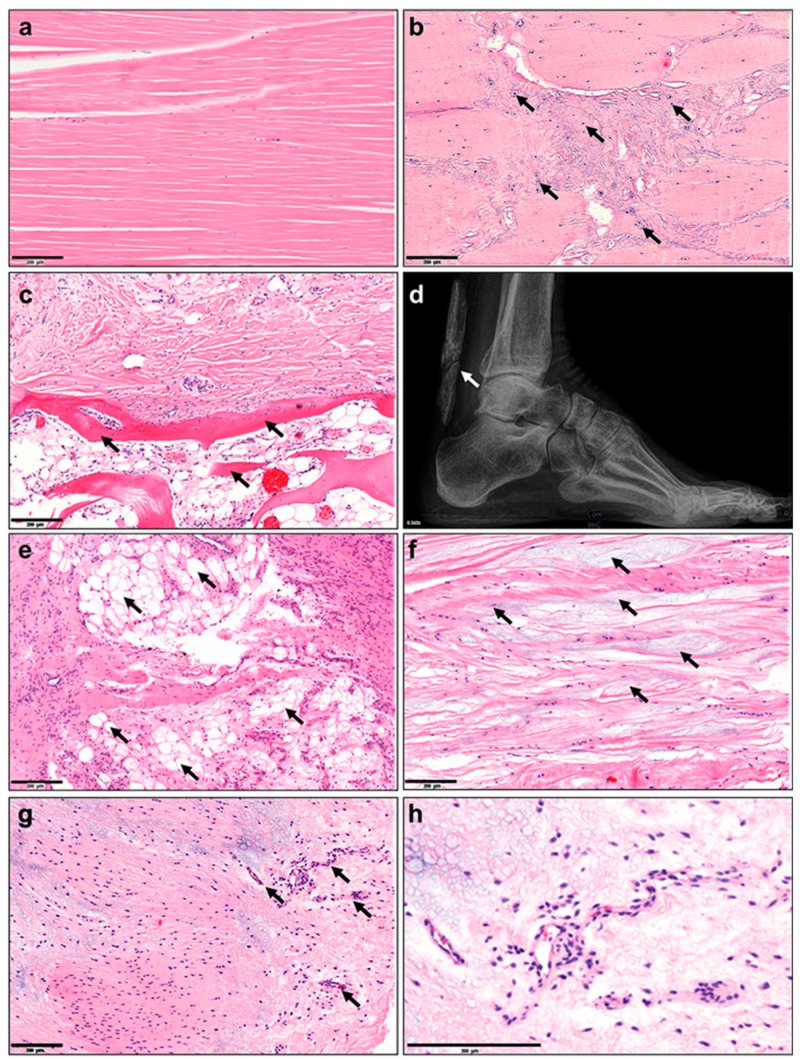
Figure 1.
Histopathological changes in tendinopathy. (a) Normal tendon tissue, (b) fiber crimping and kinking, loosening of collagenous matrix, (c) increased proteoglycan (PG)/glycosaminoglycan (GAG) production and changed cytokine profiles, (d) hypercellularity, (e) apoptosis, (f) presence of other cell types such as chondrocytes (fibrochondrogenesis), (g) osteocytes (calcification), h) adipocyte accumulation, i) hypervascularization and j) innervation. Based on Järvinen et al. [25].

