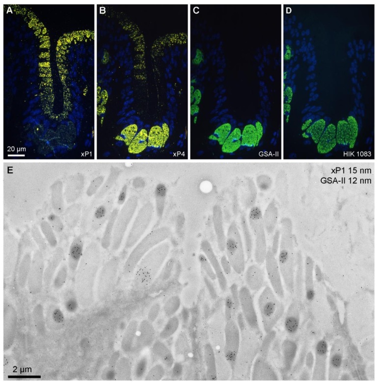
Figure 1.
Labeled ultrathin methacrylate (Lowicryl K11M) sections of X. laevis stomach fundus. Fluorescence microscopy of sequential serial sections labeled for: (A) xP1 with antiserum anti-xP1-1 (Cy3, yellow); (B) xP4 with antiserum anti-xP4-1 (Cy3, yellow); (C) mucin with GSA-II (FITC, green); (D) mucin with antiserum HIK1083 (FITC, green). DNA of the cell nuclei was stained with DAPI (blue). Scale bar: 20 µm. (E) Electron micrograph of gold labeling with the anti-xP1-1 antiserum (detected with protein A-15 nm gold particles) and mucin staining with GSA-II (12 nm gold conjugates); scale bar: 2 µm.

