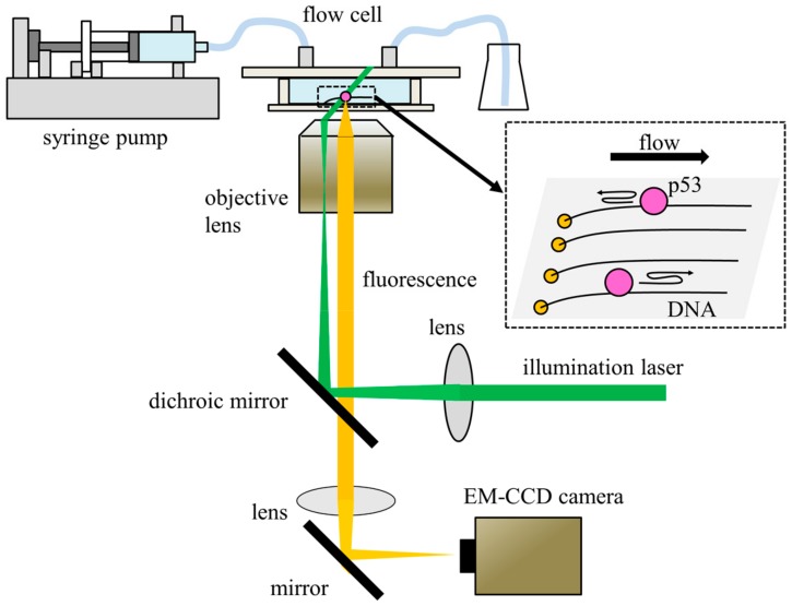
Figure 2.
Schematic diagram of representative single-molecule fluorescence microscopy. The system comprises a fluorescence microscope and a flow cell. Fluorescently labeled p53 is introduced into the flow cell using a syringe pump. DNA is tethered on one end to the MPC-polymer-coated glass surface of the flow cell in a line using the DNA garden method. The tethered DNA molecules are stretched by flow pressure. TIRF or HILO is used to illuminate DNA-bound p53 molecules. Fluorescence from the p53 molecules is detected by EM-CCD. The figure is adapted from ref. [25] with some modifications.

