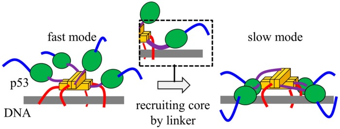
Figure 3.
Schematic diagram of the p53-DNA complex structure in 1D diffusion along a non-target DNA sequence. The linker (purple) recruits the core domain (green) to DNA (grey), triggering the conformational switch between fast and slow 1D diffusion modes. The color of p53 domains is the same in Figure 1.

