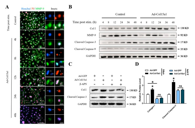
Figure 8.
Abnormal ECM remodeling persistently stimulated apoptosis of adipocytes. All groups were pretreated with PA to induce adipocytes apoptosis in (A) and (B). (A) Representative images of adipocytes morphological characteristics in different groups with the extension of stimulation time stained by Hoechst 33258 (blue), PI (red), and MMP-9 (green). Arrowheads indicate the morphological characteristics changes of adipocytes nucleus. From top to bottom represent deformed nucleus, marginalized chromatin and disintegrated nucleus, respectively. Scale bar, 100 µm (left) and 5 µm (right). (B) Relative protein levels of Col I, MMP-9, Cleaved Caspase-3 and Cleaved Caspase-9 of adipocytes in different groups with the extension of stimulation time (n = 4). (C–D) Relative protein levels of Col I and Cleaved Caspase-3 of adipocytes in different groups (n = 4). Values are presented as mean ± SEM. *p < 0.05, ** p < 0.01, ns, not significant.

