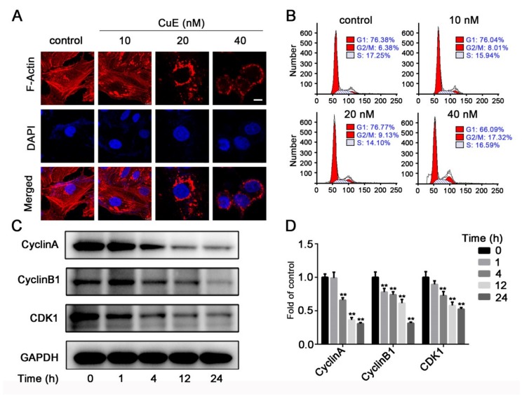
Figure 3.
(A) CuE effected cytoskeletal organization. Huh7 cells grown coverslips were treated with CuE (0, 10, 20, and 40 nM) for 6 h. Immunocytochemistry was conducted using rhodamine-conjugated phalloidin to visualize F-actin fibers (bar = 10 μm). (B) Huh7 cells were treated with CuE (0, 10, 20, and 40 nM) for 24 h. The cells were fixed and stained with propidium iodide (PI), and the DNA content was analyzed by flow cytometry. (C) Huh7 cells were treated with CuE (40 nM) for different time points (0, 1, 4, 12, and 24 h), and the protein levels of cyclin A, cyclin B1 and CDK1 were detected by Western blotting. (D) Quantitative analysis for relative protein expression levels of CyclinA, CyclinB1, and CDK1 was performed by normalizing to GAPDH. Data are mean ± SD of three independent experiments (n = 3). **: p < 0.01.

