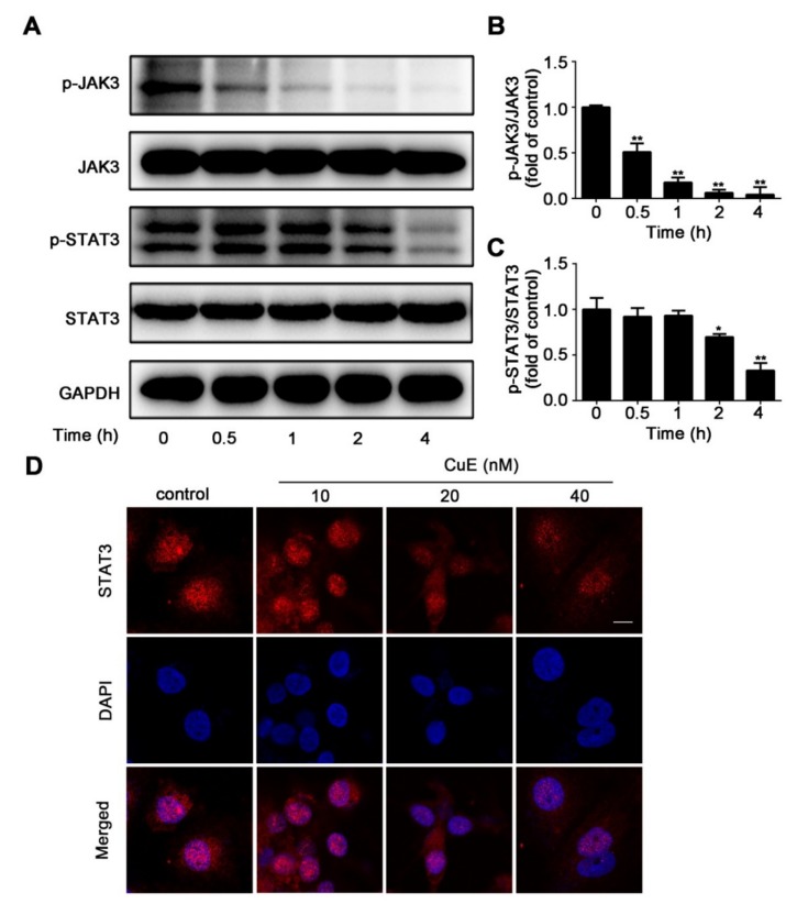
Figure 5.
CuE restrained the activation of JAK3 and STAT3 in Huh7 cells. Cells were treated with CuE (40 nM) for various hours. (A) Phosphorylations of JAK3 and STAT3 protein were determined by Western blotting. (B-C) Quantitative analysis for relative phosphorylation levels of JAK3 (B) and STAT3 (C) was performed by normalizing to the control group. Data are mean ± SD of three independent experiments (n = 3). *: p < 0.05, **: p < 0.01, compared with the control group. (D) Effect of CuE on nuclear translocation of p-STAT3 in Huh7 cells. Immunofluorescent analysis was conducted with antibody of p-STAT3 and secondary antibody conjugated to rabbit Dylight-594. Images were captured using a confocal laser scanning microscope.

