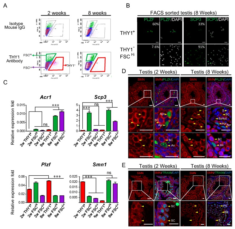
Figure 1.
The distribution of survival motor neuron (SMN) in young and adult mouse testes. (A) Fluorescence-activated cell sorting (FACS) was used to characterize and sort testicular cells from 2- and 8-week-old mice. Based on the staining intensity of Thy-1 Cell Surface Antigen (THY1) conjugated with Alexa-488 fluorescent dye, three populations were identified: SSC (THY1+), spermatocyte (THY1−FSCHi), and sperm/spermatids/spermatocyte (THY1−FSCLo). Rat Immunoglobulin G (IgG) subsequently conjugated with 488 was used as the isotype control antibody. (B) Sorted THY1+ cells from 8-week-old mice express a high percentage (60%) of promyelocytic leukemia zinc-finger (PLZF) (green, left panel), whereas THY1−FSCHi cells shows a low percentage of PLZF signal (7.6%). The meiotic marker synaptonemal complex protein 3 (SCP3) was used to characterize the THY1−FSCHi population, which mostly contains spermatocyte. The percentages of markers in different populations are indicated. (C) Determination of the expression level of Smn1 and germ cell markers in the sorted population. In the THY1+ SSC population, Smn1 showed a significantly higher expression level in 2-week-old cells (2w THY1+) but decreased to a lower level in the THY1−FSCHi spermatocyte population (2w THY1−FSCHi). In 8-week-old THY1+ SSC cells (8w THY1+), the expression level of Smn1 showed no difference compared to the spermatocyte population of 2-week-old cells but increased significantly in the THY1−FSCHi spermatocyte population (8w THY1−FSCHi). The transcript expression level of the spermatogonia-specific marker Plzf, sperm marker Acr1, and meiotic marker Scp3 showed a cell-type-dependent manner. The error bar indicates technical repeats. The expression level of each group was compared with the 2w THY1+ group. *Indicates significance, p < 0.05; **, p < 0.005. (D) Putative proliferating A-paired spermatogonia (Apr, arrowhead, upper, and middle panel) expresses the abundant amount of SMN in the testis of both 2- and 8-week-old mice, whereas undifferentiated A-single spermatogonia (As, arrowhead, lower panels) expresses PLZF (green) and possesses a lower level of SMN (red) compared with spermatocyte (SC). Sertoli cells (arrow indicated) also express a lower amount of SMN protein. (E) Validation of germ cell marker TRA98 expression in spermatocyte. SMN expressed abundantly in pachytene (PS)-stage spermatocytes colocalized with TRA98 (green); the expression decreases in round spermatocyte (RS), elongated spermatocyte (ES), and is the lowest in spermatozoa (S) in 8-week-old testis. The isotype negative control used for the double staining of antibodies is shown in Supplementary Figure S2. The scale bar in this figure represents 20 μm.

