Figure 3.
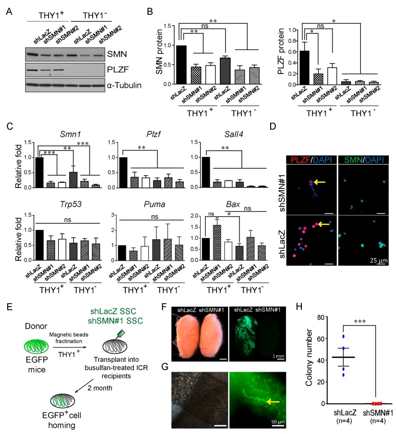
Spermatogonial stem cells (SSCs) show decreased PLZF expression and lost the ability of self-renewal after SMN knockdown. (A) Western blot analysis and its quantification (B) demonstrated decreased PLZF protein following lentiviral shSMN transduction in SSCs enriched by MACS isolated from 6–7 days postpartum (dpp) testes of C57BL/6JNarl mice (shSMN#1 and shSMN#2). Compared with the control shLacZ group, a significant difference is shown. α-Tubulin is used as the internal control of the Western blot. The original Western blot is shown in Supplementary Figure S4 (n = 2). (C) Real-time PCR analysis of SMN-depleted SSCs. Smn1, spermatogonia makers, Plzf and Sall4, and apoptotic markers Trp53, Puma, and Bax were analyzed (n = 3). (D) Immunofluorescent staining demonstrates SSCs express less PLZF in the SMN knockdown group (shSMN#1, red color, arrow indicated) compared to the control shLacZ group. 4′,6-diamidino-2-phenylindole (DAPI) is used as the counterstain (blue). (E) Schematic illustration of the process of SSC transplantation. (F) SSCs carrying enhanced green fluorescent protein (EGFP) transplanted into a recipient testis shows colonization in the shLacZ group (left testis) but not in SMN-depleted SSCs (shSMN#1, right testis). (G) Aligned EGFP-positive cells in the shLacZ control group represent the homing ability and normal proliferation (arrow). (H) Quantification of the SSC homing efficiency. Each spot indicates one replicate of the transplanted mouse. *Indicates significance, p < 0.05; **, p < 0.005; ***, p < 0.001; ns indicates no significance.

