Figure 1.
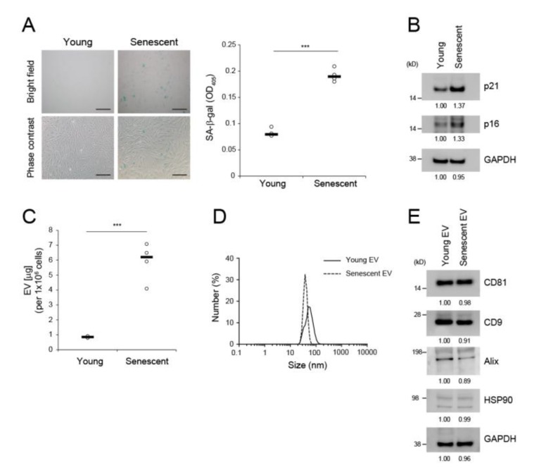
EV secretion was increased in replicative senescent dermal fibroblasts. (A) Senescence-associated (SA)-β-gal assay in young HDFs (PDL < 10) and senescent HDFs (PDL > 50). Scale bar = 50 μm. Data are means ± SD of three independent experiments on two independent senescent cell lines (*** p < 0.001). (B) Western blot analysis of p21 and p16 in young versus senescent HDFs. GAPDH was the loading control. (C) Levels of EVs derived from equal numbers of young and senescent HDFs. Protein concentrations in isolated EVs were determined by BCA assay. Data are means ± SD of four independent experiments using two independent senescent cell lines (*** p < 0.001). (D) Dynamic light scattering analysis of EVs derived from young and senescent HDFs. (E) Western blot analyses of EV markers. Five micrograms of EV protein were subjected to immunoblot analysis with anti-CD81, anti-CD9, anti-Alix, and anti-HSP90. GAPDH was the loading control.

