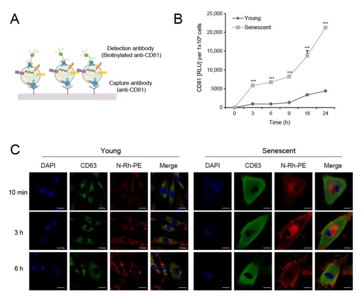
Figure 2.
EV biogenesis increased in senescent dermal fibroblasts. (A) Schematic image of sandwich ELISA using anti-CD81 antibody to detect EVs. (B) Conditioned media used to culture equal numbers of young and senescent HDFs were harvested at the indicated time points. EV levels were quantitated by sandwich ELISA for CD81. Data are means ± SD of three independent experiments using a senescent cell line (*** p < 0.001). (C) HDFs were labeled with a fluorescent lipid molecule N-Rh-PE (red) for the indicated time periods and co-stained with anti-CD63 antibody (green). Nuclei were stained with 4′6-diamidino-2-phenylindole (DAPI; blue). Fluorescence images were taken under a confocal microscope. Representative images are shown. Scale bar = 20 μm.

