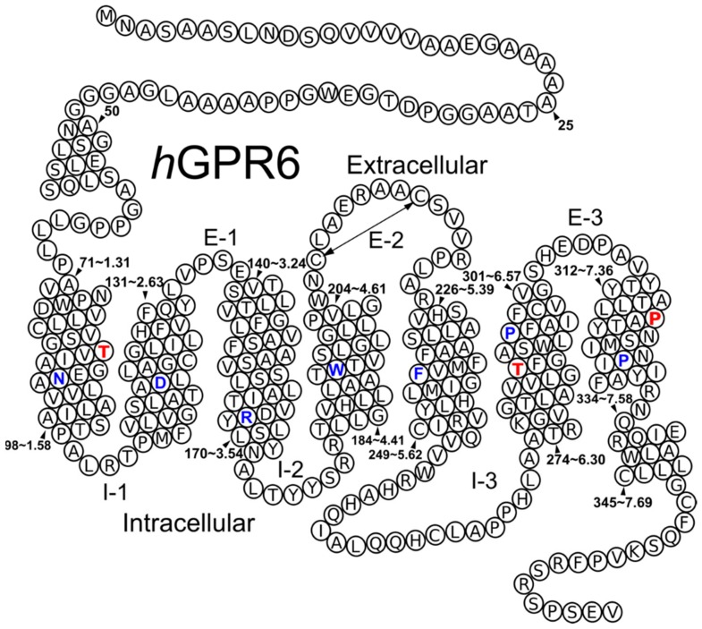
Figure 1.
Helix net representation of the human GPR6 sequence. The most highly conserved residue in each helix is colored blue. A key disulfide bridge between the extracellular loop 2 (EC-2) is indicated by a double headed arrow. Black arrowheads indicate specific residue numbers in the GPR6 sequence (absolute and Ballesteros–Weinstein numbering). The additional helix bending residues in TMH1, TMH6 and TMH7 are that were colored red.

