Figure 1.
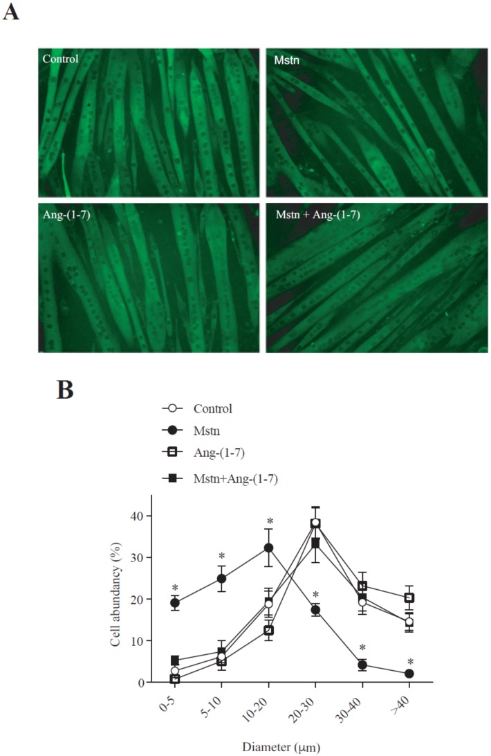
Angiotensin (1-7) (Ang-(1-7)) prevented the myostatin-induced decrease of the myotube diameter. (A) C2C12 myoblasts differentiated for five days (myotubes) were pre-incubated in the absence or presence of Ang-(1-7) (10 nM) for 60 min, and then incubated with myostatin (Mstn, 1 µg/mL) for 72 h. Myosin heavy chain (MHC; green) was detected through indirect immunofluorescence (IFI). Hoechst was used to stain the nuclei (blue). The bar scale represents 100 μm. (B) The graphics show the distribution of the myotube diameters. The values are expressed as a percentage of the total myotubes and correspond to the mean ± standard deviation (SD) from three independent experiments (n = 3; * p < 0.05 vs. control).

