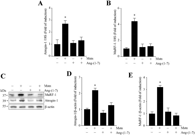
Figure 3.
Ang-(1-7) avoided the myostatin-dependent induction of atrogin-1 and MuRF-1 in the myotubes. The C2C12 cells differentiated for five days were pretreated or not with Ang-(1-7) (10 nM) for 1 h, and further with myostatin (Mstn, 1 µg/mL) for 12 or 24 h. The mRNA levels of atrogin-1 (A) and MuRF-1 (B) were determined by RT-qPCR. (C) The protein levels of atrogin-1 and MuRF-1 were detected by Western Blot analysis. β-actin was used as the loading control. The molecular weights are shown in kDa. Quantitative analysis of atrogin-1 (D) and MuRF-1 (E) levels with values normalized to β-actin. All of the values correspond to the mean ± SD from three independent experiments (n = 3; * p < 0.05 vs. control without treatment).

