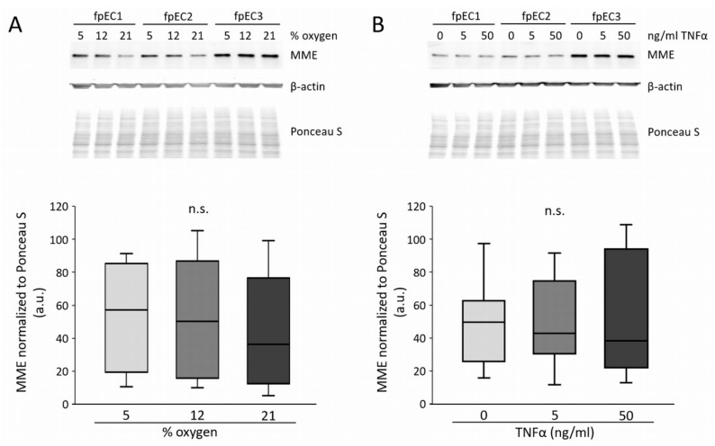
Figure 5.
Effect of oxygen and tumor necrosis factor α (TNFα) on MME protein in fpEC. (A) MME protein after culture at 5%, 12%, and 21% oxygen for 48 h. (B) MME protein after TNFα treatment (0, 5, and 50 ng/mL) for 24 h. Protein levels of β-actin were used as loading controls and protein was normalized to total protein staining with Ponceau S. Experiments were performed in n = 7 different fpEC isolations. Representative immunoblots of three fpEC isolations are shown on top. a.u.: arbitrary units; n.s.: not significant.

