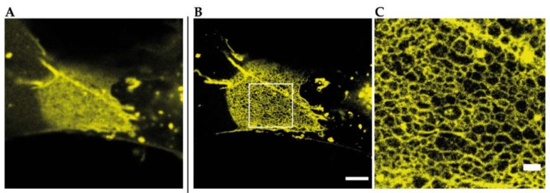
Figure 4.
Reconstituted TJ networks formed by YFP-claudin-3 in Cos7 fibroblasts (unpublished data) imaged by using STED microscopy; (A) confocal overview image of a reconstituted TJ meshwork at the overlapping area of two transfected cells; (B) STED image of the same meshwork area from A. Scale bar: 2 µm; (C) magnification of the marked area in B. Scale bar: 500 nm.

