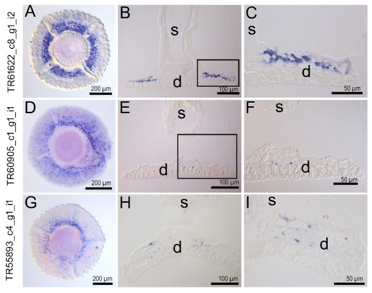
Figure A5.
Spatial distribution of labeled cells within the tube foot disc. In situ hybridization signal (blue) in a ventral view onto the tube foot disc (A,D,G), in semi-thin sagittal section though the tube foot (B, E, H). The rectangle indicated in (B) is magnified in (C), the rectangle in (E) is magnified in (F). (I) shows the lateral part of the disc of a successive section of (H). s, stem; d, disc.

