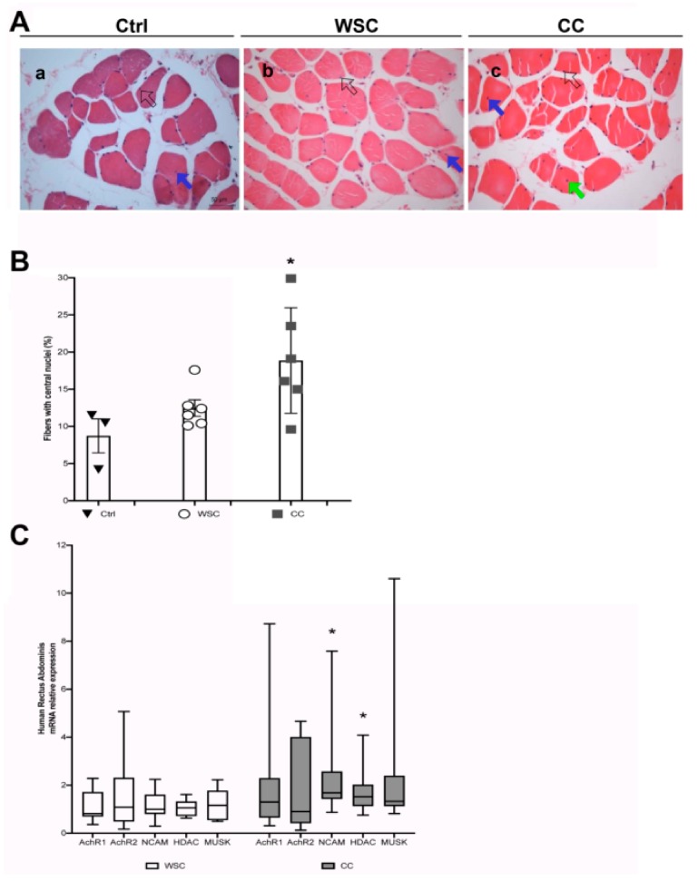
Figure 4.
An increased number of central nuclei in the human Rectus Abdominis fibers is associated with cachexia and denervation markers. (A) Representative images showing H&E staining of cross sections from the RA muscle of patients without cancer (a, Ctrl), Weight-Stable Cancer patients (b, WSC) and Cancer Cachexia patients (c, CC). Central nuclei are indicated by blue or green arrows (in the first case the nucleus is just displaced from the subsarcolemmal position, while in the second case it is closer to the center of the fiber); peripheral (i.e. normal) myonuclei are indicated by open arrows. Scale bar is 50 μm. (B) Percentage of fibers with central nuclei in muscle cross-sections of Ctrl, WSC and CC patients (a randomly chosen subset of the patients in Table 1). The three groups showed significant differences in the number of fibers with central myonuclei (F = 4.93; df = 2; p = 0.025 by ANOVA; * p < 0.05 by Tukey’s HSD test, used as a post-hoc test for CC vs Ctrl). Data are presented as mean +/- SEM, n = 3–6 for each group. (C) Q-PCR analysis on muscle from the RA of WSC (n = 10) and CC (n = 13) patients for denervation markers as indicated. Data are presented as box-and-whisker plot, showing the median +/- 10–90 percentile range, and analyzed by using Wilcoxon-Mann-Whitney test; * p < 0.05.

