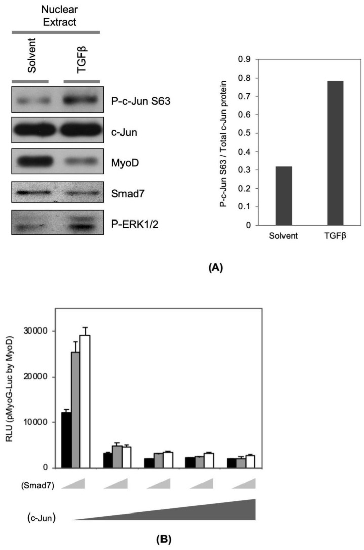
Figure 4.
TGFβ signaling modulates MyoD co-activator and co-repressor proteins. (A) C2C12 cells were seeded onto cell culture plates at equal density and maintained in TGFβ (1 ng/mL) or solvent in DM for 16 h. Nuclear protein was extracted by using NE-PER®. The amount of indicated nuclear protein was visualized with standard Western blotting technique (left panel). The band intensity of P-c-Jun and total c-Jun were measured using ImageJ, and the ratio of the P-c-Jun to the c-Jun band intensity was graphed in the presence and absence of TGFβ (right panel). (B) C2C12 cells were transfected with myogenin promoter-luciferase reporter gene construct (pMyoG-Luc) with MyoD expression vector, and increasing amounts of c-Jun expression vector (0, 0.1, 0.4, 0.8, 1.6 μg) and combinations with Smad7 expression vector (0, 0.5, and 1 μg). In addition, to monitor transfection efficiency, pCMV-β-gal construct was included in each condition. The transfected cells were maintained for 16 h in DM. Total protein samples were harvested with a luciferase lysis buffer. Luciferase activity in each condition was measured independently and normalized according to β-Galactosidase activity (n = 3, +/- SD).

