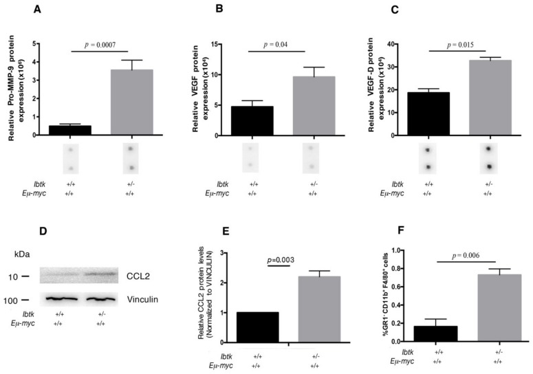
Figure 4.
Cytokine expression and recruitment of tumor-associated macrophages in the tumor lymph nodes of Ibtk+/+ Eμ-myc and Ibtk+/- Eμ-myc mice. A, B, C. Bar diagram showing the quantification of pro-MMP9, VEGF, and VEGF-D protein expression levels from Ibtk+/+ Eμ-myc and Ibtk+/- Eμ-myc cancerous mice. Densitometry data are extracted, the background was subtracted, and the data were normalized to the positive control signals, according to the manufacturer’s protocols. Values are the mean ± SEM (n = 3/genotype). Representative images of individual cytokine spots are shown from identical exposures. D. Immunoblot analysis of CCL2 and vinculin expression in the protein extracts of tumor cells from Ibtk+/+ Eμ-myc and Ibtk+/- Eμ-myc mice. Protein bands were normalized to the corresponding vinculin intensity. E. Bar diagram showing quantitation of CCL2 protein. Protein bands were measured by densitometry as arbitrary units and normalized to vinculin as the internal control. Values are the mean ± SEM (n = 3/genotype). F. Cell suspensions of tumor lymph nodes were stained with fluorescent-conjugated antibodies to reveal tumor-associated macrophages (GR1- CD11b+ F4/80+ cells) and analyzed by flow cytometry. Values are the mean ± SEM (n = 3/genotype).

