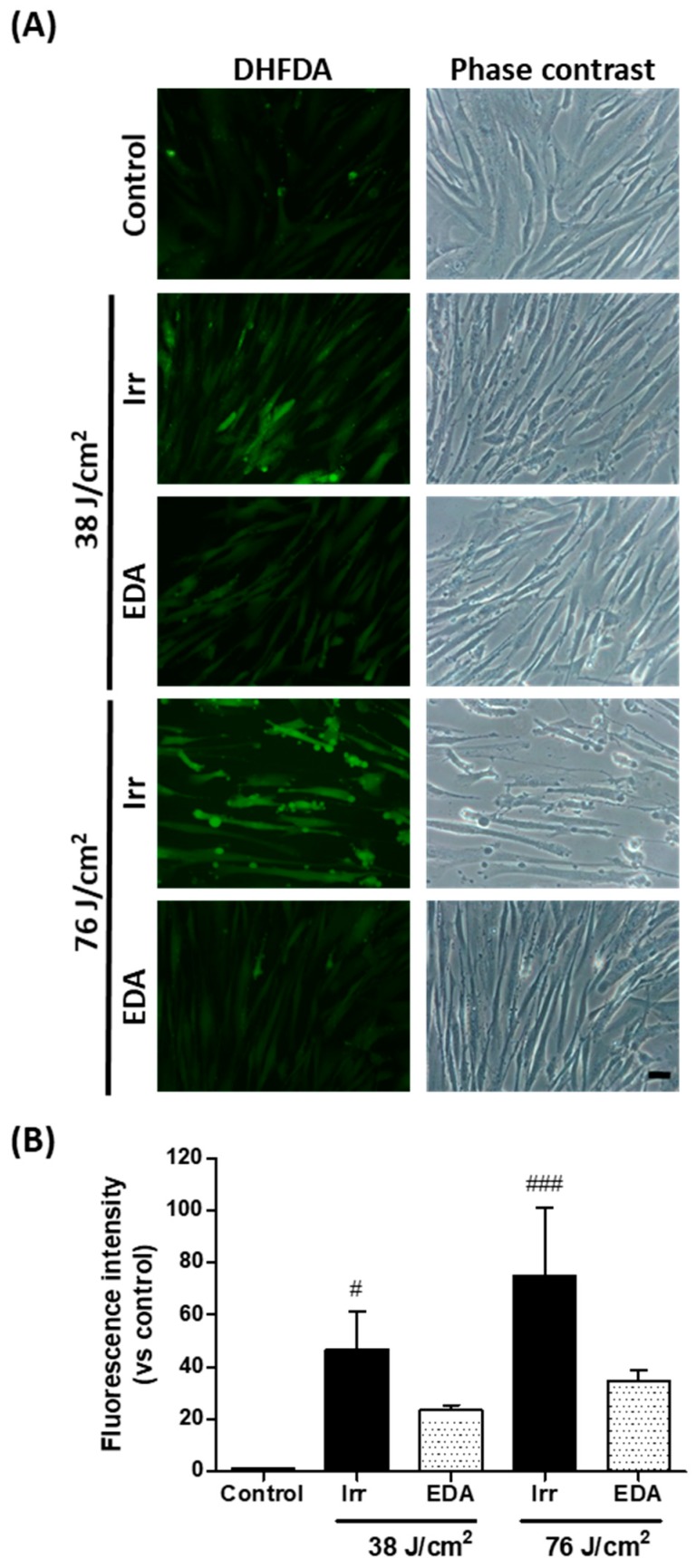
Figure 2.
Oxidative stress in human fibroblasts exposed to artificial blue light and EDA pre-treatment. Oxidative stress was evaluated by DHFDA assay. Cells were incubated with EDA 0.1 mg/mL for 24 h, loaded with DHFDA, irradiated at 38 and 76 J/cm2, washed and observed under the microscope immediately after irradiation. ROS are evidenced by green fluorescence in HDF exposed to artificial blue light and EDA (A). Quantification of DHFDA by fluorescence (n ≥ 5) (B). Data are shown as mean ±SEM. # p < 0.05, ### p < 0.001 vs control. Scale bar: 50 µm.

