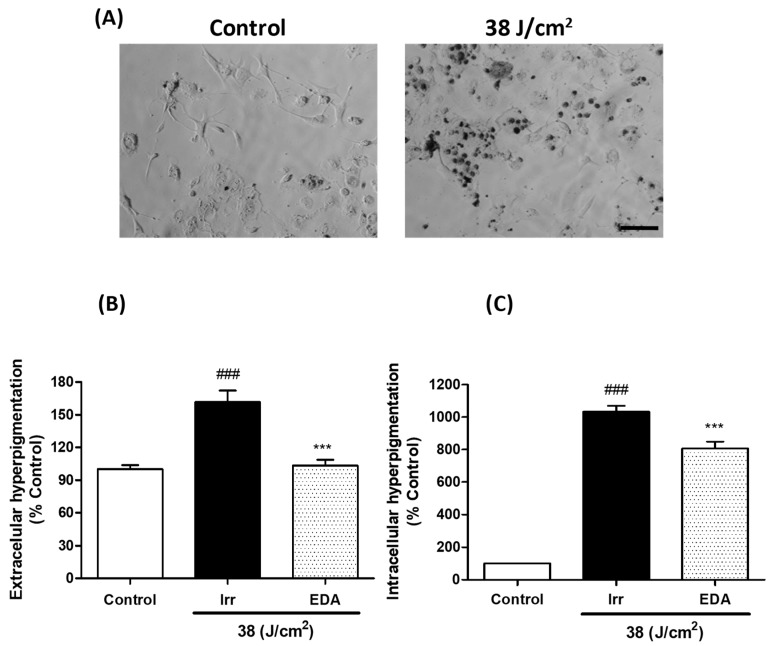
Figure 5.
Extracellular and intracellular hyperpigmentation in melanocytes exposed to artificial blue light and pre-treatment with EDA. Microphotographs show how control and irradiated melanocytes have different amounts of melanin dark granules (A). Cells were incubated with EDA 0.1 mg/mL for 24 h, irradiated (38 J/cm2), fresh medium replaced and extracellular (B) and intracellular (C) melanin pigments collected, solubilized and quantified 3 h later by absorbance measurements (n ≥ 4). Data were normalized by mg protein and expressed as % of control cells. Data are shown as mean ±SEM. ### p < 0.001 vs control; *** p < 0.001 vs irradiated. Scale bar: 50 µm.

