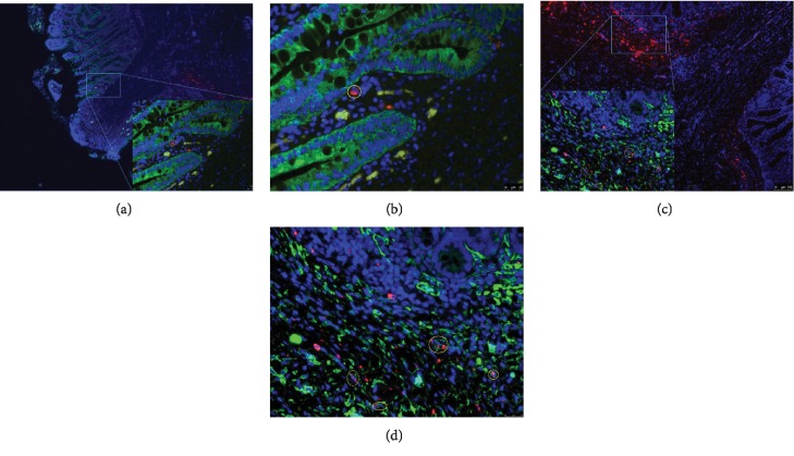
Figure 3.
MSC tracking and differentiation. (a) Tracking of grafted MSCs (50x) on d 14. (b) Tracking of grafted MSCs (400x) on d 14, showing a zoomed-in view of (a). (c) Differentiation of smooth muscle cells from grafted MSCs (50x) on d 14. (d) Differentiation of smooth muscle cells from grafted MSCs (400x) on d 14, showing a zoomed-in view of (c). Red: CM-Dil fluorescent dye. Green: anti-alpha smooth muscle actin in (a, b) and pan cytokeratin in (c, d). Blue: 4,6-diamidino-2-phenylindole.

