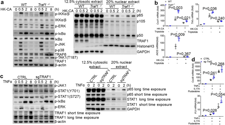
Fig. 7.
TRAF1-deficiency influences p65 in macrophages and STAT1 in keratinocytes. a. Traf1−/− and WT BMDMs pretreated with IL-4 (10 ng/ml) O/N were treated with HK-CA (MOI = 5) for indicated times. The whole cell lysates or cytosolic/nuclear extracts were prepared and probed with indicated antibodies. b. IL-4 (10 ng/ml) pretreated Traf1−/− and WT BMDMs were pretreated with triptolide (20 ng/ml) for 2 h before HK-CA (MOI = 5) treatment for 6 h. The expression of Cxcl1, iNOS, Tnfα was evaluated by qPCR. Results were reported as fold change normalized to the expression of β-actin and relative to untreated control. c. CTRL and sg-Traf1 HaCaT cells were stimulated by TNFα (20 μg/ml) for indicated times. The whole cell lysates or cytosolic/nuclear extracts were prepared and probed with indicated antibodies. d. CTRL and sg-Traf1 HaCaT cells were either stimulated or unstimulated with TNFα (20 μg/ml) plus IL17A (50 μg/ml) for 9 h, in the absence or presence of fludarabine (100 μM) for 4 h, and the expression of Cxcl8, Ilβ was evaluated by qPCR. Results were reported as fold change normalized to the expression of β-actin and relative to untreated control. For Q-PCR, data are shown as mean ± SEM and are representative of at least two experiments performed in triplicate, and were analyzed using the unpaired, two-tailed, Student’s t-test. Values of p below 0.05 represented a statistically significant difference. The Western blots are representative of at least three independent experiments

