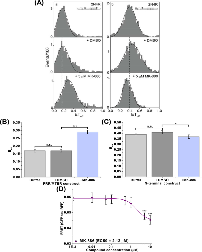
Fig. 5: MK-886 binds and perturbs tau monomer conformation.
(A) Single-molecule FRET (smFRET) measurements in the absence and presence of MK-886 with WT 2N4R tau double labeled at the proline-rich region/microtubule binding region (PRR/MTBR, left) or at the N-terminal domain (right). Tau schematic represents the labelling position for each construct. The black line is drawn from the peak of the histogram in buffer for comparison with DMSO and MK-886 samples. Representative histograms are shown. (B) Quantification of the smFRET measurements indicates that the PRR/MTBR becomes substantially more compact (increase in FRET) upon binding MK-886 (5 μM) (A, bottom left) when compared to tau in buffer (A, top left) or DMSO (A, middle left) while the N-terminal domain (C) shows only minor differences in the presence of MK-886 (5 μM) (A, right). (D) FRET analysis of the dose response of MK-886 in the cellular tau intra-molecular biosensor indicates an EC50 value of 2.12 μM, similar to that of oligomer modulation, suggesting that the change in conformational states of oligomers is due in part to conformational changes of tau monomer. Data are means ± SD of three independent experiments. *P < 0.05, ***P < 0.001 and n.s. indicates not significant by two-tailed unpaired t test.

