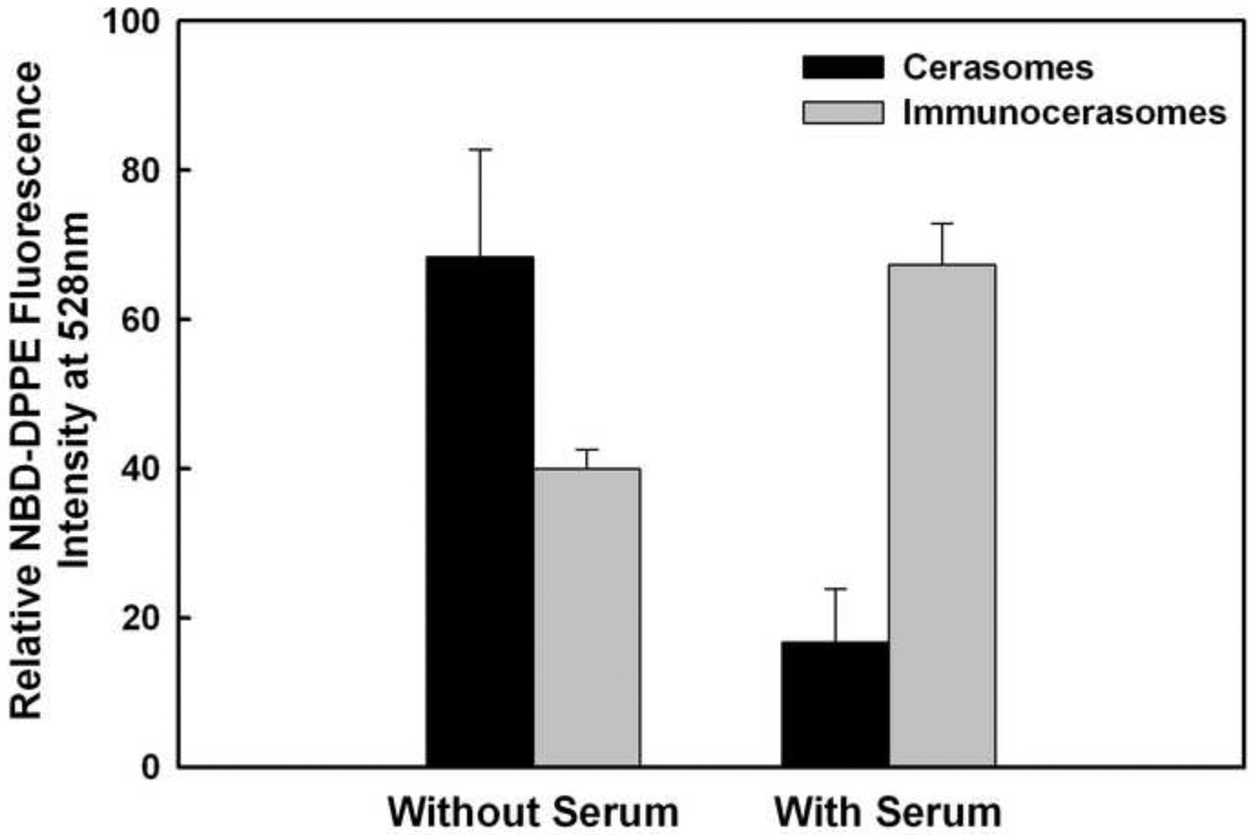
Fig. 7.

Serum effects on the cellular uptake of cerasomes and immunocerasomes by A431 cells. The cells were incubated with immunocerasomes or cerasomes at a lipid concentration of 185 μM for 3 hr. The relative amount of internalized immunocerasomes or cerasomes was measured by the NBD-DPPE fluorescence intensity of the cell lysates. The averages and standard deviation were calculated from measurement of three replicate specimens. The p values were calculated to be 0.03630 for the endocytosis of cerasomes vs. immunocerasomes in the serum-free medium, 0.00044 for the endocytosis of cerasomes vs. immunocerasomes in the serum-enriched medium 0.00610 for the endocytosis of cerasomes in serum-free vs. serum-enriched media, and 0.00277 for the endocytosis of immunocerasomes in serum-free vs. serum-enriched media, respectively.
