Fig. 3.
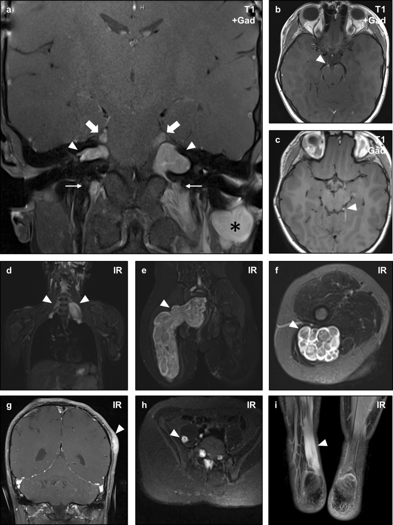
Non-vestibular schwannomas in NF2. Magnetic resonance imaging (MRI) of an NF2 patient with numerous intracranial and peripheral schwannomas. Coronal post-contrast T1-weighted sequences show bilateral vestibular (CNVIII) schwannomas (arrowheads) confirming the diagnosis of NF2, as well as bilateral trigeminal (CNV) nerve masses (large arrows), bilateral cervical nerve root masses (small arrows), and a post-auricular mass (asterisk) consistent with schwannomas (a). This patient also developed oculomotor (CNIII) schwannomas (b, arrowhead), and a small left trochlear nerve (CNIV) schwannoma (c, arrowhead). Whole body inversion recovery (IR) sequences showed large plexiform masses in the bilateral brachial plexus (d, arrowheads), and right sacral plexus (e, arrowhead). Axial slices of the latter mass demonstrate the multi-nodular plexiform architecture of the lesion (f, arrowhead). Schwannomas may also arise in unusual locations, as illustrated by a cutaneous lesion in the scalp (g, arrowhead), intramuscular lesion in the right iliopsoas muscle (h), and a diffusely infiltrative lesion in the right Achilles tendon (i, arrowhead) in this patient

