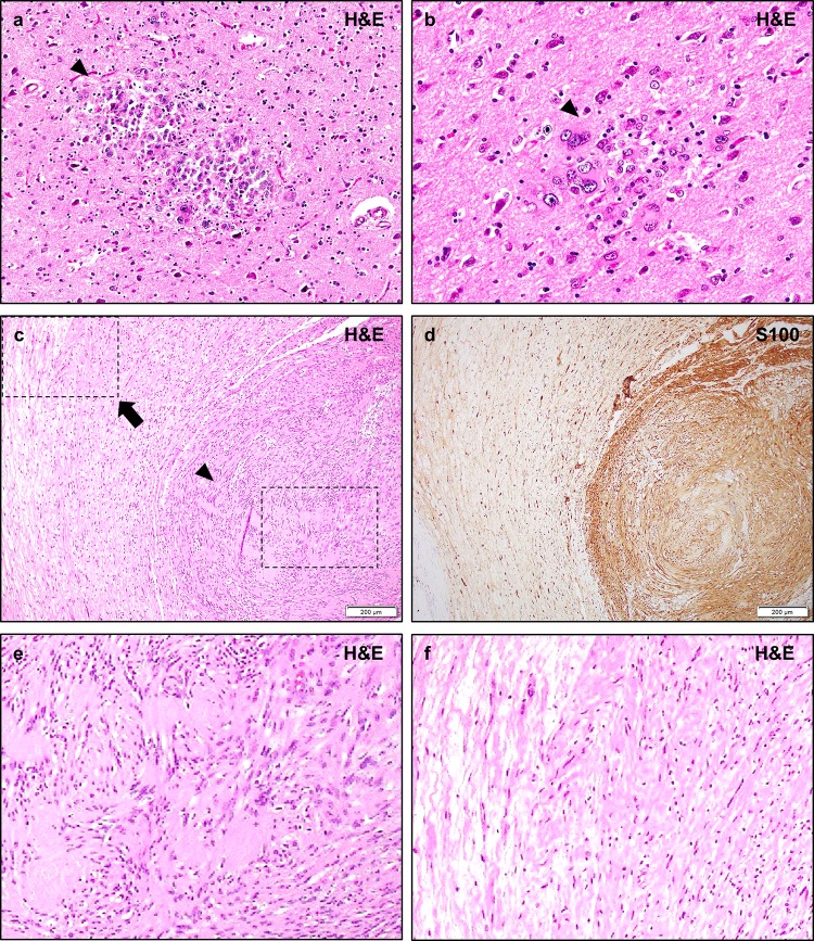
Fig. 8.
Other lesions in NF2. Post-mortem examination of NF2 patients may demonstrate small circumscribed collections of immature-appearing glial cells known as glial hamartia (a, b, arrowheads). The cells of these lesions typically exhibit eosinophilic cytoplasm, pleomorphic nuclei, and occasional multi-nucleation. The cells are strongly S100 positive, with occasional focal GFAP staining, but their precise lineage derivation is not certain. The surrounding cortex may exhibit reactive changes but does not typically exhibit features of focal cortical dysplasia. Hybrid schwannoma/neurofibromas are frequently encountered in NF2 patients (c, d), and typically exhibit nodular regions of schwannoma with typical histologic features such as Verocay bodies and hyalinized vessels, a monomorphic cellular population, and strong/diffuse S100 staining (e, arrowhead), while other regions exhibit lower density regions with mixed cellularity, loose “shredded carrot” collagen, and less prominent S100 staining consistent with a neurofibroma component (f, arrow). While pure neurofibromas are occasionally encountered in NF2 patients, they are typically sporadic solitary masses, in contrast to the multiple plexiform neurofibromas of NF1. Pathologic review shows that many lesions initially diagnosed as neurofibromas in NF2 patients are in fact misdiagnosed hybrid schwannoma/neurofibromas. Scale bars 200 μm (c, d)

