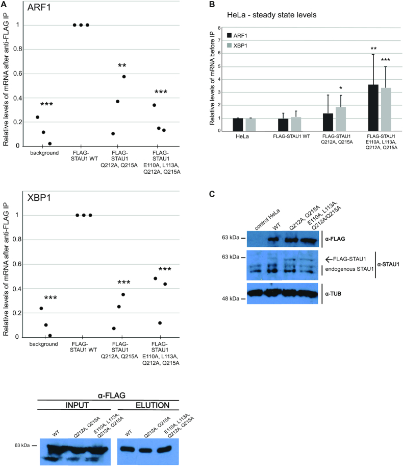
Figure 3.
Mutations in dsRBD3 and dsRBD4 impair binding to target mRNAs in vivo. (A) RT-qPCR analysis of the amount of ARF1 and XBP1 mRNAs coprecipitated with WT and mutant STAU1 from HEK293T-REx cells. The Y-axis represents the enrichment of mRNAs coprecipitated with STAU1 variants relative to the level in the whole cell lysate (input). Input ARF1 and XBP1 mRNA levels were normalized to GAPDH as an internal control. The qPCR results were analyzed by the ΔΔCt method. Scatter plot represents relative enrichments of precipitated mRNAs with FLAG-tagged STAU1 mutants relative to the FLAG-WT STAU1. Background is the level of mRNAs unspecifically bound to FLAG-beads from control HEK293T-REx where no FLAG-tagged protein was expressed. Error bars SD (n = 3–4 biological replicates), *P-value < 0.1, **P-value < 0.01, ***P-value < 0.001; P-values were calculated by two-tailed paired t-test. The western blot below the graphs shows the efficiency of FLAG immunoprecipitation. Input is the whole cell lysate before IP. (B) RT-qPCR analysis of steady-state levels of ARF1 and XBP1 mRNA in T-REx-HeLa cells upon overexpression of stably integrated STAU1 variants. Data were analyzed by ΔΔCt calculation method and normalized to GAPDH as an internal control. Bar plot represents fold enrichments relatively to control T-REx-HeLa cells. Error bars SD (n = 3 biological replicates), *P-value < 0.1; P-values were calculated by two-tailed paired t-test. (C) Western blot analysis of STAU1 protein levels upon doxycyclin induction using antibodies as indicated on the right. Tubulin was used as a loading control. Control HeLa is a cell line without any integration and expression of a FLAG-tagged protein.

