Figure 5.
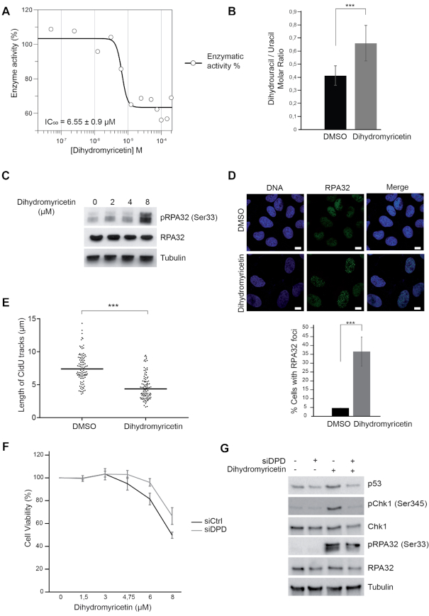
Dihydromyricetin induces DNA replication stress. (A) IC50 determination of dihydromyricetin for DHP (0.2 μM) using dihydrouracil (50 μM) as a substrate. One representative experiment is shown from three biological replicates. (B) Molar ratios of dihydrouracil versus uracil measured in U-2 OS cells treated with 20 μM dihydromyricetin for 16 h. Data from three independent biological replicates, with three technical replicates for each, are represented as mean ± S.E.M. P-values were calculated using a regression model with Poisson distribution: ***P < 0.0001. (C) Western blot analysis with the indicated antibodies of U-2 OS whole-cell extracts treated with dihydromyricetin for 48 h at the indicated concentrations. One representative experiment is shown from three biological replicates. (D) RPA32 immunofluorescence staining of U-2 OS cells treated with DMSO or 20 μM dihydromyricetin for 16 h. Bars indicate 10 μm. DNA was stained by Hoechst. Bottom panel: Histogram representation of the percentage of RPA32 foci-positive cells in a population of 100 cells. Data from three independent biological replicates are represented as mean ± S.D. P-values were calculated using a regression model with Poisson distribution: ***P < 0.0001. (E) Graphic representation of replication track lengths measured in μm (y-axis) in control and U-2 OS cells treated with 20 μM of dihydromyricetin for 16 h. The bar dissecting the data points represents the median of 100 tracts length from one biological replicate. Differences between distributions were assessed with the Mann-Whitney rank sum test. P-values: *** < 0.0001. (F) U-2 OS cells were transfected with control or anti-DPD siRNA and exposed to increasing concentrations of dihydromyricetin for 2 days. Cell viability was estimated using Cell Titer-Glo assay. Mean viability is representative of experiments performed in triplicate. Error bars represent ± S.E.M. (G) Western blot analysis with the indicated antibodies of whole cell extracts from U-2 OS transfected with anti-DPD siRNA and treated or not with 20 μM of dihydromyricetin for 24 h. One representative experiment is shown from two biological replicates.

