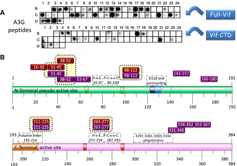
Figure 2.
Binding of full-length Vif and Vif CTD to A3G derived peptides–Peptide array screening results: (A) Binding of full length (His)6-Vif and Vif CTD 141–192 to the peptide array. Detection was performed using mouse anti-Vif monoclonal antibody followed by incubation with HRP-conjugated goat anti-mouse secondary antibody and ECL development. For more information please refer to Tables 2 and S1. (B) Schematic representation of A3G peptides that bound full-length Vif (purple boxes) and Vif CTD (red boxes) are indicated above the A3G domains.

