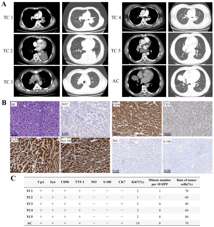
Figure 1.
Radiological and pathological results of six patients with pulmonary carcinoids. (A) Computed tomography imaging of six patients with pulmonary carcinoid. (B) Representative HE and IHC images of pulmonary carcinoids under light microscope at ×100 magnification. (C) IHC results of 6 patients with pulmonary carcinoids. IHC, immunohistochemistry; HE, hematoxylin and eosin; TC, typical carcinoid; AC, atypical carcinoid.

