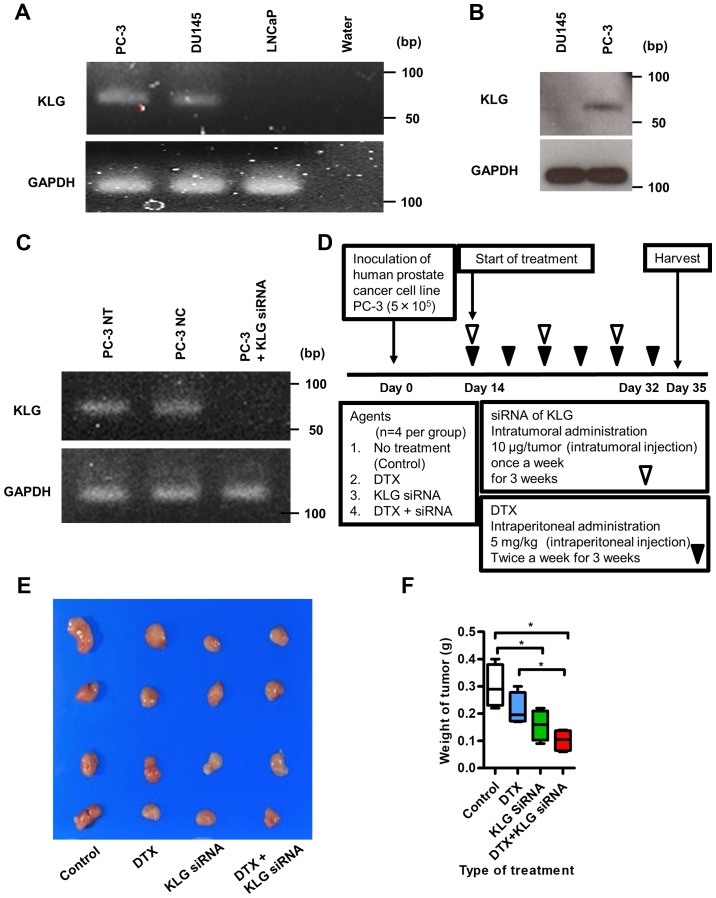
Figure 4.
Reverse transcription PCR and western blot analysis of KLG in PC-3 cells, and xenograft model. (A) Reverse transcription-PCR analysis revealed that PC-3 and DU145 cells expressed KLG RNA. (B) Western blot analysis showed that KLG protein was expressed in PC-3 cells. (C) Expression of KLG was knocked downed with KLG siRNA as shown in the reverse transcription-PCR analysis. (D) Schematic diagram illustrating the study workflow. Mice were injected with PC-3 cells (5×10⁵/tumor) together with Matrigel. After 2 weeks of inoculation, mice were randomly divided into four groups [n=4 per group; control (no treatment), DTX, KLG siRNA and DTX + KLG siRNA]. Mice were treated for 3 weeks. After 5 weeks of inoculation, mice were euthanized and xenografts were harvested. (E) Subcutaneous tumors were removed from the mouse xenograft model. (F) Tumor weight was significantly lower in the KLG siRNA and KLG siRNA + DTX treated groups than in the control and DTX only groups. Kruskal-Wallis test was conducted. *P<0.05. KLG, γ-Klotho; siRNA, small interfering RNA; DTX, docetaxel; siRNA, small interfering RNA.

