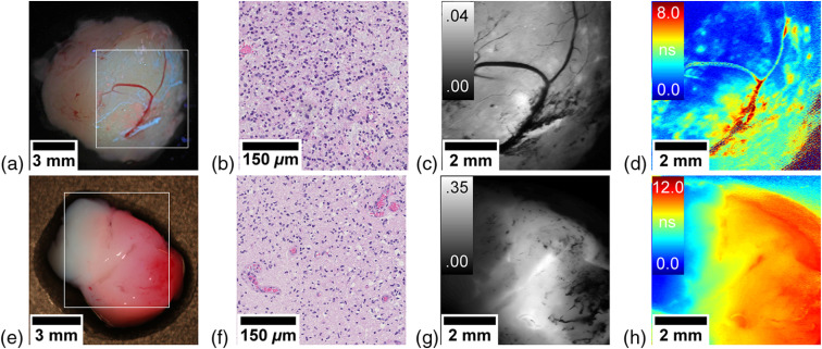
Fig. 3.
Brain tumor samples. (a)–(d) WHO grade III astrocytoma and (e)–(h) WHO grade III oligodendroglioma. Conventional white-light image by a consumer grade camera with fluorescence overlay. (a), (e) The white rectangle indicates the area of the scan; (b), (f) representative histopathological section; (c), (g) fluorescence intensity (rel.) acquired with the PMT; and (d), (h) fluorescence lifetime map (ns).

