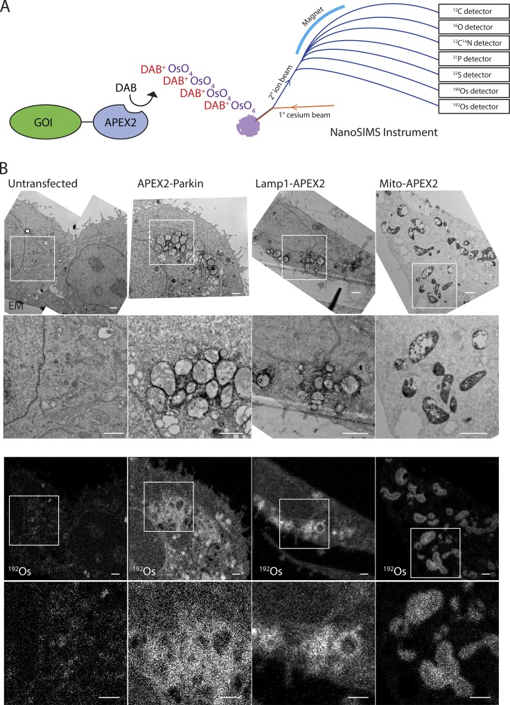
Figure 1.
Detection of APEX2 fusion proteins by MIMS. (A) Schematic depicting detection of APEX2 by the NanoSIMS instrument in parallel with ions abundant in biological samples. APEX2 fused to the gene of interest (GOI) catalyzes the polymerization of DAB, which in turn precipitates OsO4. The sample is probed with a primary beam of positively charged cesium ions. Ionized atoms and polyatomic fragments from the sample surface are focused into a secondary ion beam, which is separated in a magnetic field. Seven parallel detectors are tuned to detect specific ions of interest. (B) Adjacent ultrathin sections of HeLa cells untransfected or transfected with APEX2-Parkin, Lamp1-APEX2, or Mito-APEX2. Samples were treated with DAB and OsO4 and analyzed by TEM or MIMS for osmium ions (190Os or 192Os) or 12C14N. APEX2-Parkin and untransfected cells were treated with the mitochondrial uncoupler carbonyl cyanide m-chlorophenyl hydrazone (CCCP) 10 µM for 5 h to induce translocation of Parkin to the outer mitochondrial membrane. Representative images from a single cell are shown for each condition. These are representative of 14 untransfected HeLa cells (in two technical/biological replicates), 2 APEX2-Parkin transfected HeLa cells (in one technical/biological replicate), 11 Mito-APEX2 transfected HeLa cells (in three technical/two biological replicates), 6 Lamp1-Apex2 HeLa cells (in two technical replicates/one biological replicate). Scale bar in all images = 1 µm.

