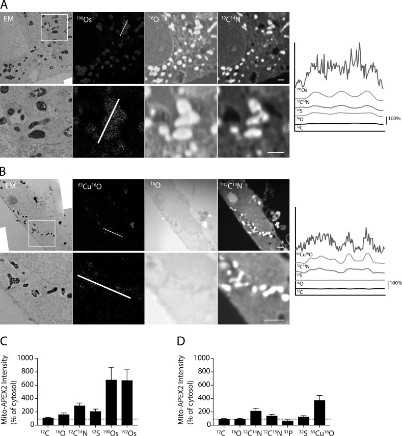
Figure 3.
CuSO4 and OsO4 enhance ionization in their vicinity by a matrix effect. (A) HeLa cell transfected with Mito-APEX2, fixed with OsO4, and imaged in adjacent ultrathin sections by EM and MIMS. Location of line scan is indicated in second image. Image is representative of 11 Mito-APEX2 transfected HeLa cells (in three technical/two biological replicates). (B) HeLa cell transfected with Mito-APEX2, stained with CuSO4 (in the absence of OsO4), and imaged in adjacent ultrathin sections by EM and MIMS. Location of line scan indicated in second image. Image is representative of three Mito-APEX2 transfected HeLa cells (in one technical/biological replicate). (C and D) Average intensity of ions from mitochondrial ROIs compared with random ROIs from the cytosol for samples fixed with OsO4 (C) or stained with CuSO4 in the absence of OsO4 (D). 14 mitochondria from one representative cell in A and B were analyzed in each condition. Dotted lines indicate 100% of cytosol intensity. Error bars represent SD. Scale bar in all images = 1 µm.

