Figure 1.
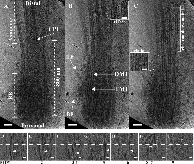
Slices through a motile cilium from respiratory epithelia. Slices at different z-heights were extracted from a tomogram to illustrate ciliary features. (A and B) Overall, the cilium base had an hourglass shape, with the TMTs of the proximal cilium giving way to DMTs (B). The basal foot (BF) and transition fibers (TF) were identified binding approximately to the TMT-containing region (B). In the DMT region, the CPC (A) and ODAs became apparent (B and insert in B). (C) The lumen of the microtubules changed along the cilium axis, from empty to striated (inset in C) to filled with electron-dense material. (D–J) We also examined the relationship between the electron-dense material inside the A-tubule and the appearance of the CPC. The dashed line in D–J denotes the start of the CPC (* in J). The CPC microtubule did not appear to be capped by a gTuRC, nor did it have any obvious attachments to the DMTs. For six of nine DMTs, we could identify the boundary between the striations and the electron-dense material (arrowheads), and in each case it was abrupt and occurred before the appearance of the CPC. Scale bars are 100 nm (A and B); 50 nm (insets and D–J).

