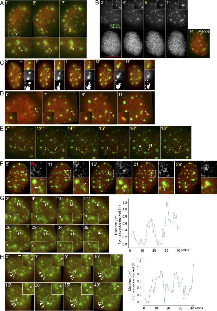
Figure S3.
Additional examples of speckle and transgene dynamics. Time stamp during HS (yellow), BAC transgene (white arrowheads, bright green), nuclear speckle (empty arrowheads, lighter green), and RNA MS2-tagged transcripts (red, mCherry-MCP). All scale bars = 1 µm. (A) Combined speckle and transgene movements after speckle movement toward and association with the transgene (category: F, Fig. 5 B). Dotted lines connect to relatively stationary speckles. (B) Movement of two speckles toward the transgene locus (category: F, Fig. 5 B). (C) Speckle protrusion toward the active transgene locus (category: speckle protrusion, Fig. 5 B), with the transgene then moving closer to speckle. (D) Speckle formation at the active transgene (white arrowhead) after HS at 7 min, with nascent transcripts appearing above background at 8 min (category: speckle formation, Fig. 5 B). (E) No persisting transcription for nonassociating locus (category: no association, Fig. 5 B). Arrow (15 min) shows direction of transgene movement. Transcriptional bursting is observed at both nonassociating transgene loci at 13 min. These bursting signals are observed again at 15 min at both loci. At 16 min, one transgene (left) associates (A) with a speckle and now maintains elevated transcript signal during the rest of the observation time, whereas the other transgene that is not associated (N) with speckle does not maintain an elevated transcript signal. (F) Coordinated movement of speckle and gene (category: coordinate movement of speckle and gene, Fig. 5 B). White arrows mark the direction of gene movements. Nuclear speckle and associated BAC transgene (white arrowhead) move together as a single unit before merging with a different speckle, suggesting a stable attachment of transgene and speckle (Video 5). (G and H) Movements of Hsp70 BAC transgene away from and back to nuclear speckles at 37°C. White arrows mark the direction of gene movements. BAC transgene shows two (G) or four (H) long-range movements away from and then back toward nuclear speckle. Category in G: FB + α, Fig. 5 B and Video 6; category in H: gene moving to a different speckle, Fig. 5 B.

