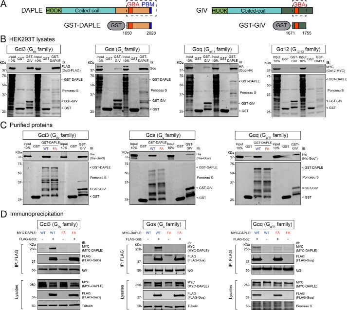
Figure 1.
DAPLE binds efficiently to Gαs and Gαq through its GBA motif. A, bar diagrams depicting the domains of DAPLE (left) and GIV (right) and the fragments of each one fused to GST used for experiments shown in this figure. B, DAPLE binds efficiently to Gαi3, Gαs, and Gαq but not to Gα12, whereas GIV only binds efficiently to Gαi3 among the G proteins tested. Lysates of HEK293T cells transfected with Gαi3–FLAG, Gαs, Gαq–HA, and Gα12–MYC were incubated with GST, GST–DAPLE, or GST–GIV immobilized on GSH-agarose beads. Bead-bound proteins were detected by Ponceau S staining or IB as indicated. C, DAPLE WT but not DAPLE F1675A (FA) binds to purified Gαi3, Gαs, and Gαq. His–Gαi3, His–Gαs, or His–Gαq* were incubated with GST, GST–DAPLE (WT or FA mutant), or GST–GIV immobilized on GSH-agarose beads, and bead-bound proteins were detected by Ponceau S staining or IB as indicated. D, DAPLE WT, but not DAPLE FA mutant, co-immunoprecipitates with Gαi3, Gαs, and Gαq. Lysates of HEK293T cells co-expressing full-length MYC–DAPLE (WT or FA mutant) with the indicated FLAG-tagged G proteins (or no tagged G protein as negative control) were subjected to IP with a FLAG antibody, and bound proteins were detected by IB as indicated. The lower immunoblot panels (lysates) correspond to aliquots of the starting material used for IPs shown in the upper panels (IP: FLAG). All results presented in this figure are representative of at least three independent experiments (n ≥3).

