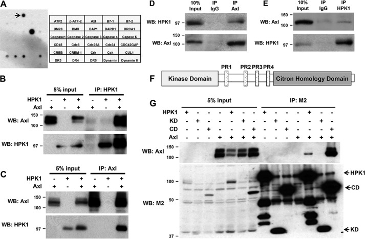
Figure 1.
HPK1 physically interacts with AXL. A, antibody array results and map showed that HPK1 interacts with AXL (marked by arrow). B and C, interaction between HPK1 and AXL was detected by reciprocal co-immunoprecipitation. HEK293T cells were transfected with pCMV-AXL alone or co-transfected with pCMV-AXL and Flag-HPK1 expression plasmids, and co-immunoprecipitation was performed as described under “Materials and methods.” D and E, endogenous HPK1 interacted with endogenous AXL in Jurkat cells. F, a schematic drawing of the domain structure of HPK1 protein. G, AXL binds to the C-terminal domain of HPK1. HEK293T cells were transfected with AXL alone or co-transfected with AXL and Flag-HPK1, Flag-HPK1 KD, or Flag-HPK1 CD expression plasmids. Cell lysates were collected for co-immunoprecipitation with M2 beads. Immunoprecipitated (IP) AXL was detected by immunoblotting. WB, Western blotting.

