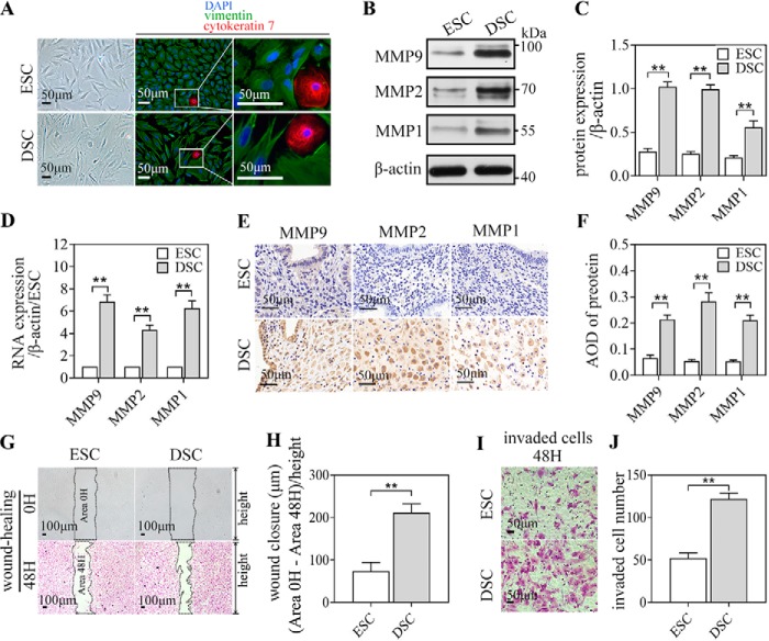
Figure 1.
MMP1, MMP2, and MMP9 expression, migration, and invasion ability of ESCs/DSCs. A, primary stromal cells isolated from secretory endometrium (n = 3∼5/group) and early pregnancy decidua (n = 3∼5) were identified as stromal cells based on their fusiform appearance, observed via an optical microscope and immunofluorescence staining: vimentin-positive (mesenchyme origin–specific, green) and cytokeratin 7–negative (epithelial cell–specific, red) DAPI, 4,6-diamidino-2-phenylindole. These isolated cells were analyzed via Western blotting and quantitative real-time PCR to investigate the expression of MMP1, MMP2, and MMP9. B, and C, Western blot of ESCs/DSCs and its quantitative representation. D, relative mRNA expression. E and F, immunocytochemistry of the secretory endometrium and decidua against MMP1, MMP2, and MPP9 and quantitative representation of the average optical density (AOD) of each protein. Primary ESCs and DSCs were subject to wound healing and an invasion assay in vitro to explore their migration and invasion ability. G, photographs recorded at 0 and 48 h following application of the wound. The length of wound closure is equal to the difference in wound area divided by the height of the wound after converting to micrometers by scale. H, quantitative representation. I and J, the invaded cells in the invasion assay were recorded photographically after 48 h (I), and the quantitative representation of the number of invaded cells is shown (J). **, p < 0.01; error bars represent standard error of the mean. The data presented are from three independent experiments.

