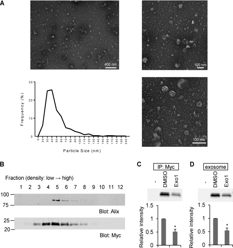
Figure 2.
The Nogo-A C-terminal region is enriched in exosomes. A, negative stain transmission EM images of the exosome fraction derived from HEK293T cell culture medium, with particle diameter distribution represented in a histogram. n = 546 particles. B, culture medium from Nogo-A–Myc–transfected HEK293T cells was separated by sucrose density centrifugation. Gradient fractions from the top were immunoblotted with anti-Alix and anti-Myc antibodies. C and D, HEK293T cells were transfected with vector (−) or Nogo-A–Myc. 24 h after transfection, media were changed to DMSO or Exo1 containing medium and cultured for another 12 h. Then the culture supernatants were immunoprecipitated (IP) with anti-Myc antibody or exosome-fractionated. Mean ± S.E., n = 3 independent experiments. *, p < 0.05; Student's two-tailed t test.

