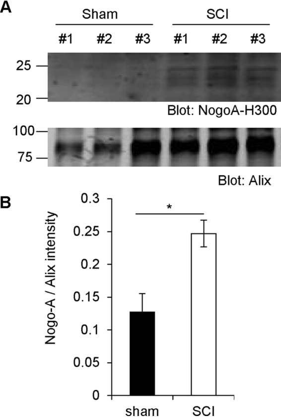
Figure 6.

Increased Nogo-A fragment levels after spinal cord trauma in vivo. A, WT mice had their spinal cord crushed and were sacrificed 3 days after surgery. The spinal cords were then taken out and homogenized in TBS. Exosome fractions were immunoblotted with anti-Nogo-A and anti-Alix antibodies. B, quantification of Nogo-A divided by Alix intensity. Mean ± S.E., n = 3 animals. *, p < 0.05; Student's two-tailed t test.
