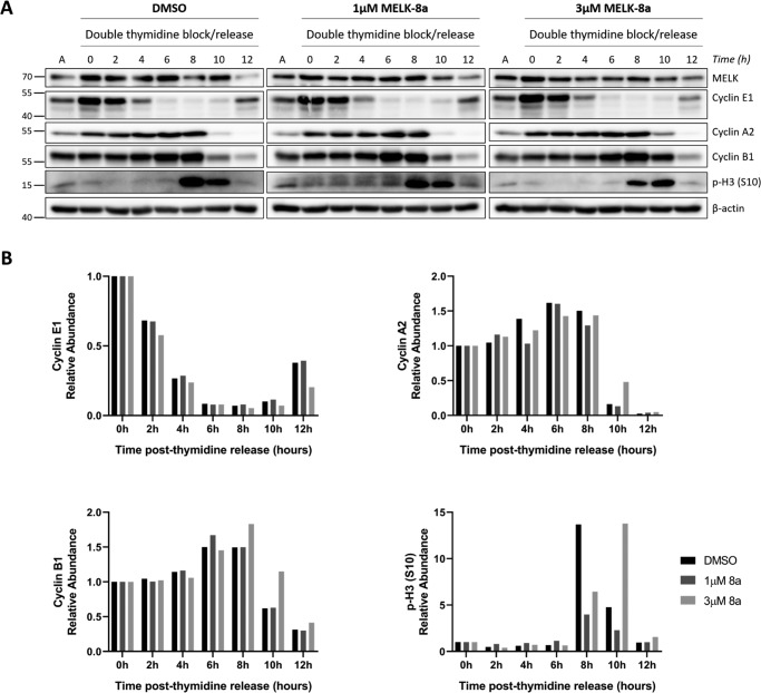
Figure 4.
MELK inhibition with 3 μm 8a causes delayed mitotic entry. A, HeLaS3 cells were synchronized at G1/S with double-thymidine block and released into DMSO or 1 or 3 μm 8a. Cells were collected every 2 h over a 12-h time course. Cells were lysed and immunoblotted with the indicated antibodies. The splits in the image indicate that the samples from DMSO and 1 and 3 μm 8a conditions were immunoblotted separately due to space constraints. Asynchronous HeLaS3 lysate was loaded in the lanes labeled A to normalize among the blots. The data shown here are representative of three independent experiments. B, the signal intensity of the cyclin E1, A2, and B1 and p-H3 (Ser-10) bands was quantified for the replicate shown in A using Bio-Rad Image LabTM software. The ratio of the quantified signal intensity at each time point relative to the 0-h time point was computed for each condition. The resulting ratios are plotted as bar charts for each protein, with 1.0 indicating no change relative to the 0-h time point.

