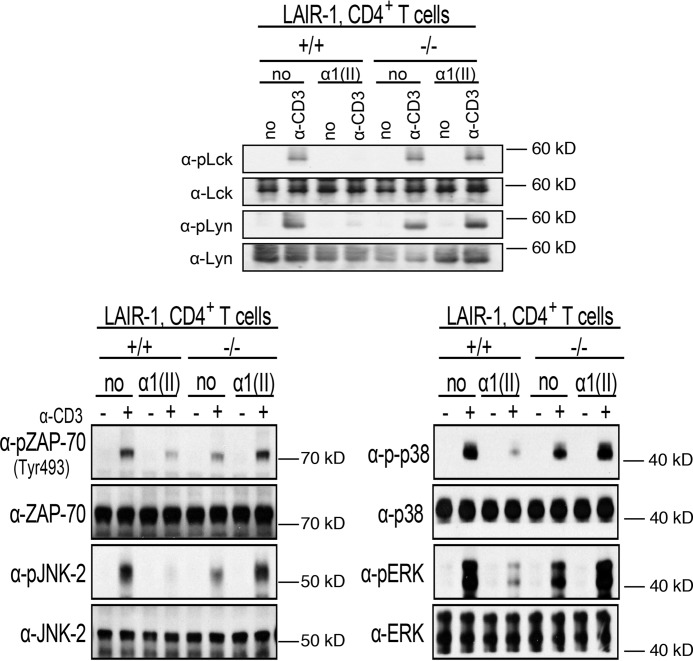
Figure 1.
Inhibition of murine T-cell receptor signaling by α1(II) in CD4+ T cells isolated from WT or LAIR-1-deficient mice. Naive CD4+ T cells were isolated by negative selection as described under “Experimental procedures” and pretreated with α chains of type II collagen (α1(II)) (200 μg/ml) overnight prior to stimulation with α-CD3. LAIR-1 expression peaked after 18 h of culture with collagen. Cells lysates were collected and separated using SDS-PAGE gels. The separated proteins were then electrotransferred onto nitrocellulose membranes and analyzed for phosphorylation of the indicated proteins by Western blotting. The upper panel shows activation of the Src kinases Lck and Lyn. The lower panels show activation of ZAP-70 (pZAP-70–Tyr-493), as well as the MAP kinases JNK (pJNK-2), p38 (pp38), and ERK 1/2 (pERK). The membrane was stripped and reblotted with non–phospho-specific antibodies. This figure is a composite of several individual gels and is representative of three separate experiments.

