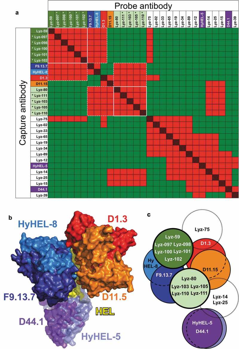
Figure 4.

Epitope mapping of anti-HEL antibodies. (a) Competition patterns of anti-HEL clones tested. Red and red/black cross-hatched squares indicate no binding of the probe antibody. Green squares indicate binding of the probe antibody. Mouse antibodies of known epitope specificity are highlighted in shades of blue, orange, red and purple. Anti-HEL clones selected based on VH/VL pairing are shown with asterisks. White lines and dotted lines indicate cross-competition with other clones in parallel lineages and mouse anti-HEL antibodies. (b) Composite structure of HEL bound to reference mouse antibodies (PDB 1YQV, 1FDL, 1FBI, 1MLC, 1NDG and 1JHL). Variable regions of antibodies are color-coded as in panel a. (c) Graphical representation of epitope groups of antibodies in parallel lineages, mouse anti-HEL antibodies and additional selected rat anti-HEL antibodies. Antibodies and parallel lineages colored as in panels a and b.
